EQUIPMENT
The Bioimaging Centre from INEB has currently the following equipment available:
1. Spectral Confocal Microscope Leica TCS-SP5 AOBS.
2. Confocal Raman Microscope, LabRAM HR800 UV, Horiba Jobin-Yvon.
3. Imaging Flow cytometer ImageStreamX.
4. Atomic force microscope
5. Inverted Fluorescence Microscope/ Coupling Stage
6. In vivo micro-ultrasounds imaging, Vevo 2100 (230 V).
1. Spectral Confocal Microscope Leica TCS-SP5 AOBS.
Our Leica TCS SP5 inverted confocal microscope provides a full range of scan speeds at the highest resolution. With three detectors and working in the AOBS (Acousto Optical Bream Splitter) mode, we can acquire bright, noise-free images with minimal photo damage at high speed.
Our system allows: 3D imaging, colocalization, excitation fingerprinting, FRET, FRAP and FLIP, RICS, photoswitching and photoactivation. Live cell imaging experiments can also be performed as the equpiment can control temperature and CO2.
Configuration: lasers available: 405, 488, 561, 594 and 633 nm; objectives: 10X dry, 20X immersion, 40X and 63X oil and 63X glicerol; adapters for 35 mm in diameter plates, well plates and 25 mm in diameter coverslips; 3 detectors: 1 Hybrid detector and 2 PMTs; motorized stage. In addition, a workstation for the analysis of the data is available.
2. Confocal Raman Microscope, LabRAM HR800 UV, Horiba Jobin-Yvon.
This equipment allows chemical imaging in unlabeled samples, providing information, in a non-destructive manner, of the structure and chemistry of not only materials but also biological samples with a resolution down to the optical diffraction limit (~ 200 nm).
Our system allows for spectra collection in 2D and 3D. Thanks to the confocal setup, detailed images and analysis can be obtained and fluorescence interference can be reduced.
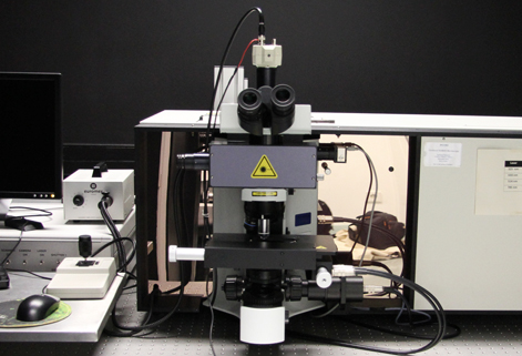
Configuration: lasers: 325, 514, 633 and 785 nm; objectives: 10x, 50X and 100X detector: CCD air cooled; spectrograph with gratings: 600, 1800 and 2400 l/mm and software for data acquisition and analysis: LabSpec 5.
3. Imaging Flow cytometer ImageStreamX.
This equipment combines the speed, fluorescence sensitivity and statistical strength of a flow cytometer with a high resolution microscopy system, giving a picture of every event. The high versatility of the analysis software allows for multiple data analysis. Our system allows the acquisition of six different channels:one for brightfield images, one for sidescatter images and four additional for different fluorochromes in use.
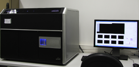
The ImageStreamX is equipped with a 488 and a 788 nm lasers. Objective: 40X. The table of suggested fluorochromes for being used in our ImageStreamX can be seen here. In addition, a workstation for the analysis of the data is available.
4. Atomic force microscope.
This technique provides 3D topographic images and surface roughness, at the nanometric scale, as well as adhesion force determination using Force Spectroscopy.
The equipment has two multipurpose scanners for AFM with 10 and 100 µm2, and performs in the following modes: Contact, Non-Contact, and Intermittent-contact (Tapping™ mode and Magnetic Acoustic Mode - MAC™). The MAC Mode™ is generally recognized as the AFM “gentle” mode, and consists in a magnetic cantilever directly controlled by a magnetic field. It integrates the oscillating mode (Acoustic AC) and the phase imaging (digital) as well. This mode is indicated for measurements in fluids, being characterized by low contact forces applied in extremely delicate samples, such as: proteins, DNA, RNA, cells and other biological structures.
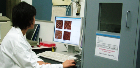
The available equipment is capable of performing measurements both in air and in liquid (a specific liquid cell is available).
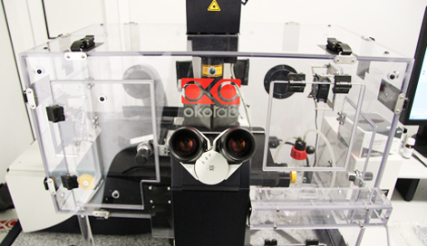
5. Inverted Fluorescence Microscope/Coupling Stage, Zeiss Axiovert 200M.
Inverted fluorescence microscopes allow for live-cell imaging applications and are capable of producing fluorescence illumination through an episcopic and optical pathway.
Our systems is equipped with a motorized stage and chamber for temperature and Co2 control. The Axiovert 200M is equipped also with the moduls: Timelapse, Z-Stack, Mark&Find, MosaiX , AutoMeasure and AutoMeasure Plus.
Configuration: fluorescence filters for: 365, 470, 565 and 640 nm; objectives: 5X air, 10X air, 20X air, LD, 40X air, LD, 40X oil, 63X oil, 63X LD, 100X oil; adapters for 35 mm in diameter plates and adapter for well plates.
6. In vivo micro-ultrasounds imaging, Vevo 2100 (230 V).
For non-invasive real-time in vivo micro-imaging of small animal research. It combines ultra-high frequency with a high-resolution digital imaging platform. It enables the simultaneous and in real-time acquisition of anatomical, functional, physiological and molecular information down to 30 µm.
Our system allows working in M-mode, PW Doppler Mode and Color Doppler Mode. Image analysis for physiolical trace including ECG, respiration waveform and body temperature.
The Vevo2100 is equipped with a MS400: 38Hz Microscan transducer with a broadband frequency: 18 MHz – 38MHz. Capable of working in M-mode, PW Doppler Mode and Color Doppler Mode. Image station plus imaging station extension with injection mount. Also the software package can be used for B-mode (2D) image capture and analysis, Cineloop image review, capture and display with integrated physiolical trace including ECG, respiration waveform and body temperature.