Past Meetings
High Throughput Screening and Image Analysis for BioSciences . 18th - 22nd June 2018
Fundamentally practical, this course aims at introducing the participants in experimental design, image acquisition, image and data analysis for high throughput experiments. Essential skills when generating massive amount of imaged-based data in the biosciences field. Lectures will be given by specialist in the field and participants will learn how to use state of the art technology for high throughput (HT) experimentation and open-source software for image and data analysis. Attendants will get acquainted with use of automated liquid handling equipment and high content (HC) imagers such as IN Cell Analyzer (HCS microscope) and ImageStream (imaging flow cytometer). Interaction with HTS/HCS specialists will be fostered due to the restricted amount of participants.
----------------------------------------------------------------------------------------------
Workshop in Atomic Force Microscopy coupled to Inverted Fluorescence Microscopy
Atomic Force Microscopy (AFM) has revealed as a powerful tool to study human pathology, in fields ranging from cancer, cardiovascular and blood diseases, since it is suitable to perform studies on different molecules, cell/ tissue types at physiological conditions. The main principle is based in the interatomic forces stablished between a probe tip and the sample causing the cantilever to deflect as the samples’ surface topography changes. A laser light reflected from the back of the cantilever measures the deflection of the cantilever. In this lab session the participants will be introduced to the AFM/IFM techniques, having the opportunity to learn about the determination of morphometric parameters to the characterization of biomechanical properties of cells. As well, students will learn how to use different software for the analysis of the data arising from these studies. Applets software will be used to analyze the Thermal tune data to obtain the force constant of the cantilevers and to analyze AFM force distance curves, to calculate the mechanical properties of the samples.

----------------------------------------------------------------------------------------------
Hands on workshop on Image Analysis for Imaging Flow Cytometry . 7th-9th June 2017
Essentially practical, this course aims at introducing the students in the use of the imaging flow cytometer and image data analysis. This hands-on course is divided in two main parts covering from the use of the imaging flow cytometer (Imagestream) and basic image analysis (Part A) to advanced image analysis focusing on machine learning for Imagestream data using the Cell Profiler environment and batch analysis with ImageJ amongst others (Part B).
Imaging flow cytometry is a state of the art technology that combines the statistical strength, sensitivity, and high speed of a conventional flow cytometer by rapidly acquiring large number of cells with the quantitative image analysis of the detailed cellular imagery with morphologic (and functional) information captured by a microscope. Similar to flow cytometry, fluorescence intensity and cell complexity can be analyzed, but furthermore and amongst others, morphological characterization, protein translocation and colocalization, can also be evaluated. The high versatility of the analysis software allows for the quantification of a diverse set of parameters granting the user complete autonomy in regard to the features to be included in the data analysis. The second part of this workshop will cover machine learning for Imagestream data using the Cell Profiler environment, and batch analysis and script writing as well as several plugins for the solution of common issues using ImageJ.
.........................................................................................................................................................................
COURSE: Introduction to data analysis and image processing with MATLAB
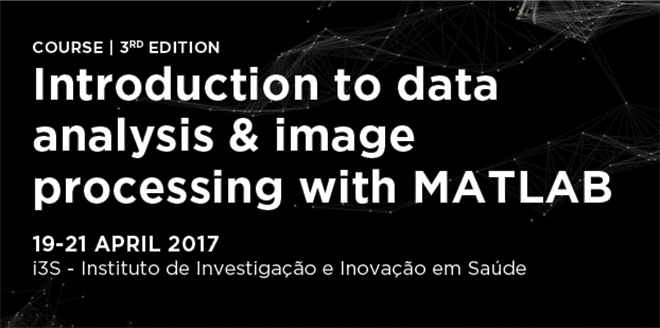
Quantification is an essential component in every scientific domain. And innovation frequently falls on the ability to extract more information from the same experimental data.
This is an introductory “hands on” course where the concepts and techniques will be explored directly in the MATLAB environment. MATLAB is a well-established tool in scientific and technical computing, with key advantages such as being extremely versatile/powerful and easy to learn. This course is especially suited for people engaged in research wanting to start using MATLAB in the context of scientific data analysis and image processing (quantification).
Participants are invited to bring their own data and an analysis problem of interest to be addressed in the course (as a project), with the help of the tutors.
.........................................................................................................................................................................
BIOIMAGING 2016. 5th INTERNATIONAL SYMPOSIUM IN APPLIED BIOIMAGING
DISEASE IN FOCUS
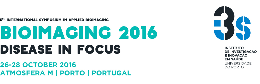
This year the Symposium, designed as a forum for those interested in the application of bioimaging tools for life and medical sciences, will run under the motto “Disease in Focus”. As is our goal, the work of our invited speakers and of all participants covers a wide range of topics – in one meeting, and in a unique style, we put together researchers that don not commonly meet. Following on the success of the previous editions, the Symposium this year is again preceeded by hands-on workshops covering state-of-the-art techniques/analysis in bioimaging.
......................................................................................................................................................................
COURSE: Introduction to data analysis and image processing with MATLAB
Quantification is an essential component in every scientific domain. And innovation frequently falls on the ability to extract more information from the same experimental data.
This is an introductory “hands on” course where the concepts and techniques will be explored directly in the MATLAB environment. MATLAB is a well-established tool in scientific and technical computing, with key advantages such as being extremely versatile/powerful and easy to learn. This course is especially suited for people engaged in research wanting to start using MATLAB in the context of scientific data analysis and image processing (quantification).
..........................................................................................................................................................................
Hands on workshop on Image Analysis for Imaging Flow Cytometry
Essentially practical, this course aims at introducing the students in the use of the imaging flow cytometer and image data analysis. This hands-on course is divided in two main parts covering from the use of the imaging flow cytometer (Imagestream) and basic image analysis (Part A) to advanced image analysis focusing on machine learning for Imagestream data using the Cell Profiler environment and batch analysis with ImageJ amongst others (Part B).
Imaging flow cytometry is a state of the art technology that combines the statistical strength, sensitivity, and high speed of a conventional flow cytometer by rapidly acquiring large number of cells with the quantitative image analysis of the detailed cellular imagery with morphologic (and functional) information captured by a microscope. Similar to flow cytometry, fluorescence intensity and cell complexity can be analyzed, but furthermore and amongst others, morphological characterization, protein translocation and colocalization, can also be evaluated. The high versatility of the analysis software allows for the quantification of a diverse set of parameters granting the user complete autonomy in regard to the features to be included in the data analysis. The second part of this workshop will cover machine learning for Imagestream data using the Cell Profiler environment, and batch analysis and script writing as well as several plugins for the solution of common issues using ImageJ.
.........................................................................................................................................................................
AFM BioMed Conference Porto 2016
AFM BioMEd Conference is dedicated to life sciences and nanomedicine applications and will investigate how Atomic Force Microscopy (AFM) solves relevant biological bottlenecks and provides innovative solutions for healthcare. The conferences link international academica and industrial experts in life sicence, being involved either with AFM use or with medical/biological studies. Te meetings provide a clear comprehensive overview on advances in this field through keynote presentations, lectues, poster sessions and discussion forums. Topics included single molecules, membrane, and cell biology and nanomedicine studies combining imaging and affinity measurements. Presentations stress the impact of AFM techniques in life sciences and nanomedicine. The meetings act as forum to promote innovative cross linked research and highlight the power of integrating AFM with other optical and analytical techniques.
....................................................................................................................................................
BIOIMAGING 2015. 4th INTERNATIONAL SYMPOSIUM IN APPLIED BIOIMAGING
THE PRE-CLINICAL CHALLENGE IN 3D
This year the event took place in Porto on November 5-6, under the topic – The pre-clinical challenge in 3D. A hands-on workshop on bioimaging with four different topics preceded the symposium (November 4th). Both the symposium and the workshop took place in a very informal environment.
The symposium was designed as a forum for those interested in the application of bioimaging tools for life and medical sciences, covering aspects ranging from imaging modalities to analysis and simulation.
We have introduced this year a grand CHALLENGE focused on the on automatic classification of tumor malignancy in the context of breast cancer.
For more information please visit the symposium website.
....................................................................................................................................................
BIOIMAGING 2014. 3rd INTERNATIONAL SYMPOSIUM IN APPLIED BIOIMAGING.
CAPTURING LIFE IN A PIXEL
In the follow up of the success of the previous Symposia in Applied Bioimaging that were organized during the last two years in Porto, the Bioimaging Centre for Biomaterials and Regenerative Therapies at INEB organized, in collaboration with IBMC and IPATIMUP the 3rd edition of this series of symposia.
In 2014 the event took place in Porto on October 16-17, under the topic – Capturing life in a pixel. A hands-on workshop on bioimaging with four different topics preceded the Symposium (October 15). Both the Symposium and the workshop took place in a very informal environment.
The Symposium was designed for those who have an interest in the application of bioimaging, from the fields of Bioimaging applied to Biomaterials and Regeneration through integrative approaches from molecular to biomedical imaging.
For more information please visit the Symposium website.
…………...…………..…………..…………..…………..…………..…………..……..………..…………..…………..
WORKING WITH IMAGEJ AND ICY:
HANDS ON IMAGE ANALYSIS FOR MICROSCOPY
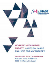
This hands-on workshop was directed to users working on biology and biomedicine that require objective analysis of microscopy image data. ImageJ, its distribution Fiji and ICY are widely-used public-domain software for microscopy image analysis. ImageJ/FIJI are based on an open source architecture, which enables functionality extensibility using plugins and Macros. ICY is an open source software that is compatible with ImageJ plugins but which enables other functionalities.
In this context, we were focused on the basic operation of both ImageJ/FIJI and ICY as well as several plugins for the solution of common issues in image analysis such as segmentation, particle analysis, co-localization, deconvolution, spot count and particle tracking. A tutorial on ImageJ/FIJI plugin programming also took place together with scripting and graphical programming for ICY.
…………...…………..…………..…………..…………..…………..…………..……..………..…………..…………..
BIOIMAGING 2013 | 2nd INTERNATIONAL SYMPOSIUM IN APPLIED BIOIMAGING
3-4 OCTOBER 2013
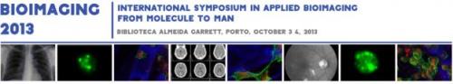
In the follow up of the success of the 1st Symposium in Applied Bioimaging that was organized last year in Porto, the Bioimaging Centre for Biomaterials and Regenerative Therapies at INEB, launched, in collaboration with IBMC and IPATIMUP the 2nd edition of this series of symposia.
This year the event took place in Porto on October 3-4, under the topic - From Molecule to Man.
The Symposium was designed for those who have an interest in the application of bioimaging, from the fields of molecular to biomedical imaging.
…………...…………..…………..…………..…………..…………..…………..……..………..…………..…………..
BIOIMAGING 2012. 1st INTERNATIONAL SYMPOSIUM IN APPLIED BIOIMAGING
19-21 SEPTEMBER 2012
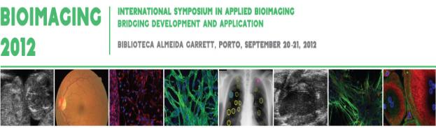
In the follow up of the establishment of the Bioimaging Centre for Biomaterials and Regenerative Therapies at INEB this symposium was launched, in collaboration with IBMC and IPATIMUP, as the first of a series of symposia in Applied Bioimaging. The topic of the 1st edition of the Symposium was “Bridging Development and Application”.
The course was designed for those who have an interest in the application of bioimaging, especially in the field of biomaterials, tissue engineering and regenerative medicine.